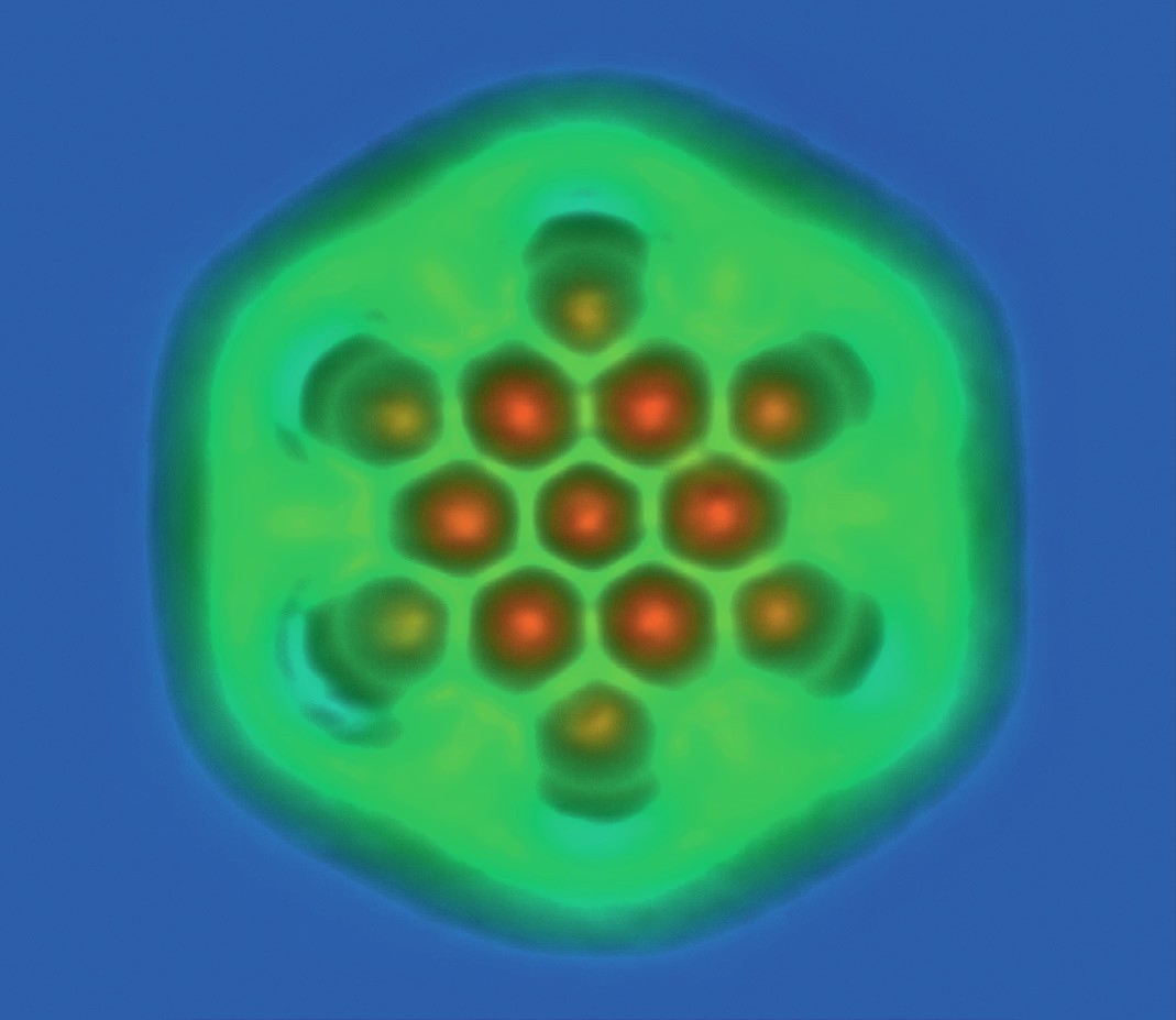
Less than a decade ago, the arrangements of atoms in molecules could not be seen directly, but physics-based techniques are now changing this.
In atomic force microscopy (AFM, Figure 1) a probe with a single-atom tip is scanned slowly across a surface, and the resulting tiny movements are detected by a laser beam and used to build up an image (Figure 2). AFM can be used to make images of individual molecules. Figure 3 shows a hexabenzocoronene (HBC) molecule, which is formed from six benzene rings (C6H6) and has a diameter of 1.4nm. The HBC, which is being studied as a potential nanomaterial, was made at Centre National de la Recherche Scientifique (CNRS) in Toulouse, France, and imaged using an AFM by IBM researchers.
Your organisation does not have access to this article.
Sign up today to give your students the edge they need to achieve their best grades with subject expertise
Subscribe


