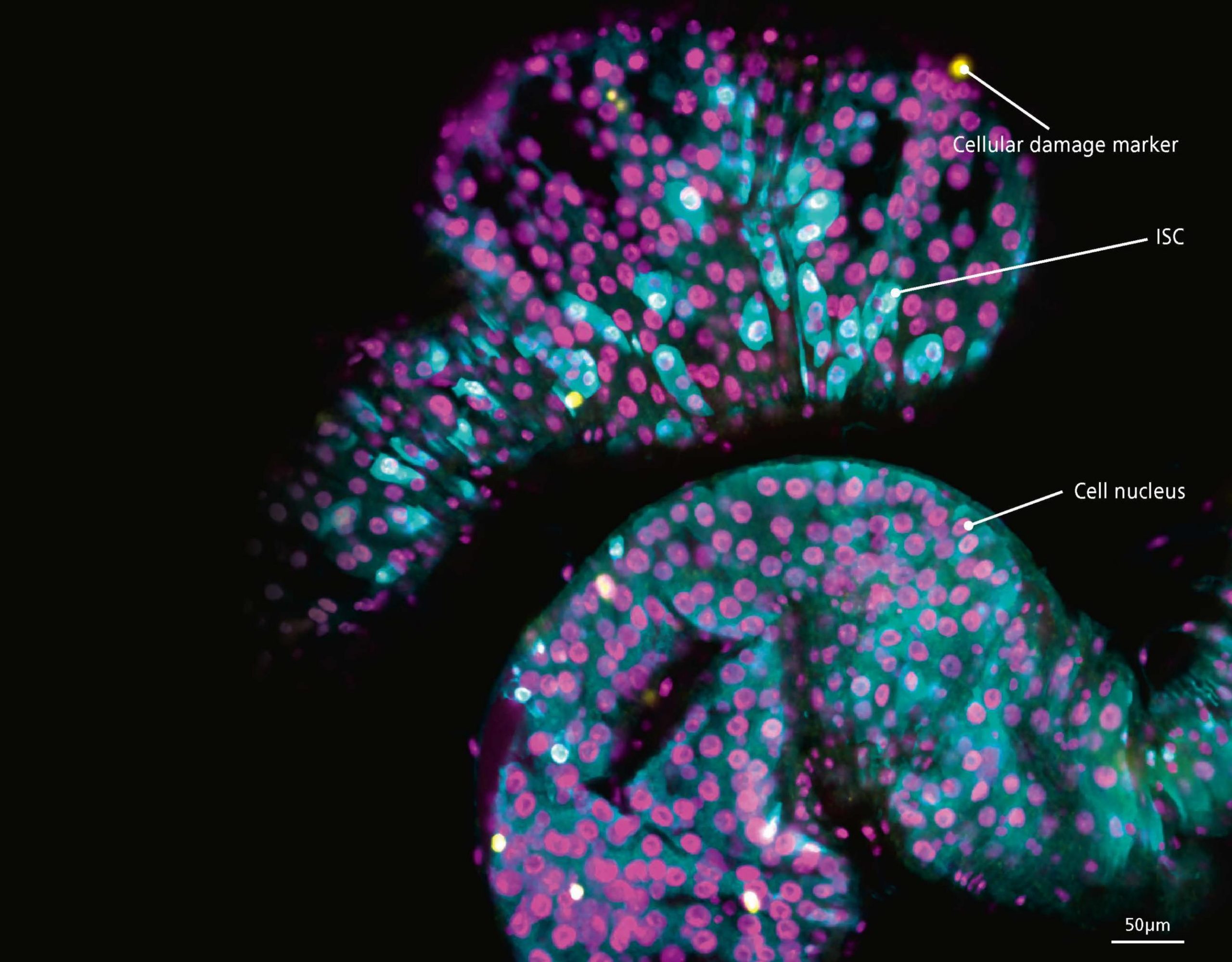
Our research uses the fruit fly, Drosophila melanogaster, as a model organism. We are studying gut diseases, including colorectal cancer. A healthy gut comprises a range of different cell types. The intestinal stem cells (ISCs – cyan fluorescence in the image above) are involved in replenishing and regenerating the intestine if it is damaged. Different genetic tools allow us to create models of gut diseases in Drosophila that help us to study the origins and mechanisms of the diseases.
The image was taken using a confocal microscope. It shows cells in the gut of a fly in which we had increased the expression of an oncogene that triggers the development of gut tumours. The ISCs divide by mitosis, proliferate and migrate from their boundaries in response to the damage. The increase in proliferation mimics the development of a tumour (tumourigenesis). The yellow dots indicate division of, and damage to ISCs.
Your organisation does not have access to this article.
Sign up today to give your students the edge they need to achieve their best grades with subject expertise
Subscribe


